在儿童恶性肿瘤中, 淋巴瘤的发病率仅次于白血病和颅内肿瘤, 是儿科最常见的恶性实体瘤, 预后与组织病理类型有关。儿童恶性淋巴瘤主要以非霍奇金淋巴瘤(non-Hodgkin lymphoma, NHL)为主, 约占70%~80%。儿童NHL则以Burkitt' s淋巴瘤和淋巴母细胞淋巴瘤、弥漫性大B细胞淋巴瘤等病理类型为主, 恶性程度常较高, 易出现全身播散和侵袭。越来越多的研究发现, TOPK/PBK(T-LAK cell-originated protein kinase/PDZ-binding kinaze)是T-LAK细胞来源的蛋白激酶, 是介导T-LAK细胞和T细胞的细胞毒功能的重要信号, 在多种恶性肿瘤如乳腺癌、小细胞肺癌、食管癌、胃癌、结直肠癌、宫颈癌等高表达[1-5]。TOPK/PBK致瘤机制复杂, 主要通过参与调控肿瘤细胞周期、促进肿瘤细胞增殖、抑制细胞凋亡以及促使肿瘤侵袭和迁移, 导致肿瘤发生发展。而TOPK/PBK与淋巴造血系统肿瘤的关系国内外报道较少。本研究通过免疫组化检测恶性淋巴瘤与淋巴结反应性增生患儿的淋巴结标本中TOPK/PBK的表达, 探讨TOPK/PBK与儿童恶性淋巴瘤的关系, 以及与病理类型和临床特征间的关系, 为儿童淋巴瘤的靶向治疗提供研究基础。
1 资料与方法 1.1 研究对象以湖南省人民医院2005~2015年经病理科确诊的80例恶性淋巴瘤患儿(霍奇金淋巴瘤12例、非霍奇金淋巴瘤68例)为研究对象, 并以20例淋巴结反应性增生患儿作为对照, 实验组、对照组年龄均为0~14岁。淋巴瘤的诊断及分类参照WHO造血与淋巴组织肿瘤病理学与遗传学分类标准[6]进行。
1.2 免疫组织化学方法检测TOPK/PBK表达取淋巴瘤及淋巴结反应性增生患儿的淋巴结石蜡组织标本进行切片, 脱蜡、水化后将切片置于EDTA抗原修复缓冲液(pH9.0)中进行抗原修复, 然后置于3%过氧化氢溶液中阻断内源性过氧化物酶, BSA封闭后滴加约50 μL TOPK/PBK一抗(浓度1 mg/mL), 湿盒内4℃孵育过夜后加入50 μL Anti-rabbit二抗(浓度1 mg/mL), 室温孵育50 min。DAB显色, Harris苏木素复染细胞核, 脱水封片, 显微镜镜检并采集图像进行分析。用已知TOPK/PBK表达阳性的淋巴瘤切片做阳性对照, 阴性对照则用PBS缓冲液替代一抗。
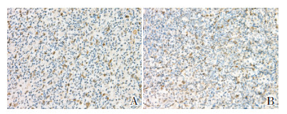
阳性结果判断:TOPK/PBK阳性表达呈棕黄色颗粒, 主要定位于细胞质, 少数定位于细胞核。在不同部位分别取5个高倍视野(×400), 每个视野记数200个肿瘤细胞, 计算阳性细胞百分率。
1.3 统计学分析采用SPSS 13.0软件进行数据处理。计数资料采用率表示, 多组间比较采用X2检验, 两两比较采用X2分割检验。P < 0.05为差异有统计学意义。
2 结果 2.1 淋巴瘤和淋巴结反应性增生患儿的TOPK/PBK阳性表达情况淋巴瘤和淋巴结反应性增生患儿的细胞质和少数细胞核内均可见清晰的浅黄或棕黄色颗粒状信号, 以淋巴瘤患儿的阳性率高于淋巴结反应性增生, 见表 1、图 1。
| 表 1 淋巴瘤和淋巴结反应性增生患儿TOPK/PBK的阳性表达情况 [例(%)] |
|
|

|
图 1 淋巴瘤和淋巴结反应性增生患儿的TOPK/PBK表达(10×40) 淋巴瘤患儿(A)及淋巴结反应性增生患儿(B)淋巴结石蜡切片的细胞质和细胞核内均可见TOPK/PBK颗粒状浅黄或棕黄色阳性表达 |
80例淋巴瘤中霍奇金淋巴瘤12例:结节性淋巴细胞为主型2例、经典型霍奇金淋巴瘤10例(结节硬化型2例、混合细胞型6例、富于淋巴细胞型与淋巴细胞消减型各1例); 非霍奇金淋巴瘤68例:淋巴母细胞淋巴瘤20例、成熟B细胞淋巴瘤19例、成熟T/NK细胞淋巴瘤13例、未分型16例。见表 2。
| 表 2 80例儿童淋巴瘤病理类型情况 [例(%)] |
|
|
HL、NHL的TOPK/PBK阳性表达率的差异无统计学意义(P > 0.05), 见表 3。
| 表 3 霍奇金淋巴瘤及非霍奇金淋巴瘤的TOPK/PBK阳性表达率比较 [例(%)] |
|
|
68例非霍奇金淋巴瘤中, 明确了病理亚型的52例非霍奇金淋巴瘤中TOPK/PBK阳性的30例(58%), 三种不同类型NHL的TOPK/PBK阳性率以淋巴母细胞淋巴瘤最高, 成熟B细胞淋巴瘤与成熟T/NK细胞淋巴瘤的阳性率差异无统计学意义(P > 0.016)。见表 4。
| 表 4 TOPK/PBK在不同类型非霍奇金淋巴瘤的阳性表达 |
|
|
TOPK/PBK主要通过激活MAPK信号通路, 导致MAPK家族p38MAPK、ERK、JNK1突变, 引起机体内一系列癌基因或转录因子如c-Myc、Sap-1、Elk-1、c-fos表达上调, 参与调节细胞增殖, 抑制凋亡和分化, 诱导肿瘤的发生[7]。本研究发现儿童恶性淋巴瘤的TOPK/PBK阳性率高于淋巴结反应性增生, 与Simons-Evelyn等[8]、Nandi等[9]研究相符。
本研究还发现, TOPK/PBK阳性率在HL与NHL之间的差异无统计学意义; 而NHL不同亚型间, 具有高度侵袭性的淋巴母细胞淋巴瘤的TOPK阳性表达率高于成熟B细胞、T/NK细胞淋巴瘤。提示TOPK/PBK表达与NHL的恶性程度相关, 肿瘤恶性程度越高, TOPK/PBK阳性表达率越高。刘宇宏等[10]研究也表明, TOPK/PBK在正常人外周血和增生的扁桃体B细胞中表达较弱, 而在一些Burkitt淋巴瘤细胞中高表达, 在免疫母细胞(活化的B淋巴细胞)淋巴瘤、弥漫大B细胞淋巴瘤的表达比惰性淋巴瘤高。因此, 根据TOPK/PBK表达水平可能推测NHL类型或者恶性程度。
综上所述, TOPK/PBK在儿童恶性淋巴瘤的淋巴结组织中表达上调, 通过TOPK/PBK的表达水平可能预测NHL的病理类型或者恶性程度, 并可能为儿童淋巴瘤的治疗提供新的靶点。
| [1] |
Ramirez-Avila L, Garner OB, Cherry JD. Relative EBV antibody concentrations and cost of standard IVIG and CMV-IVIG for PTLD prophylaxis in solid organ transplant patients[J]. Pediatr Transplant, 2014, 18(6): 599-601. DOI:10.1111/petr.12315 (  0) 0) |
| [2] |
Raab-Traub N. Novel mechanisms of EBV-induced oncogenesis[J]. Curr Opin Virol, 2012, 2(4): 453-458. DOI:10.1016/j.coviro.2012.07.001 (  0) 0) |
| [3] |
Hall LD, Eminger LA, Hesterman KS, et al. Epstein-Barr virus:dermatologic associations and implications:part Ⅰ.Mucocutaneous manifestations of Epstein-Barr virus and nonmalignant disorders[J]. J Am Acad Dermatol, 2015, 72(1): 1-19. DOI:10.1016/j.jaad.2014.07.034 (  0) 0) |
| [4] |
Iwakiri D. Mechanisms of Epstein-Barr virus-mediated oncogenesis[J]. Gan To Kagaku Ryoho, 2015, 42(10): 1133-1136. (  0) 0) |
| [5] |
赖辉红, 马廉. EB病毒相关恶性肿瘤研究进展[J]. 中国小儿血液与肿瘤杂志, 2013, 18(6): 245-249. (  0) 0) |
| [6] |
Arber DA, Orazi A, Hasserjian R, et al. The 2016 revision to the World Health Organization classification of myeloid neoplasms and acute leukemia[J]. Blood, 2016, 127(20): 2391-2405. DOI:10.1182/blood-2016-03-643544 (  0) 0) |
| [7] |
Vogelstein B, Papadopoulos N, Velculescu VE, et al. Cancer genome landscapes[J]. Science, 2013, 339(6127): 1546-1558. DOI:10.1126/science.1235122 (  0) 0) |
| [8] |
Simons-Evelyn M, Bailey-Dell K, Toretsky JA, et al. PBK/TOPK is a novel mitotic kinase which is upregulated in Burkitt's lymphoma and other highly proliferative malignant cells[J]. Blood Cells Mol Dis, 2001, 27(5): 825-829. DOI:10.1006/bcmd.2001.0452 (  0) 0) |
| [9] |
Nandi A, Tidwell M, Karp J, et al. Protein expression of PDZ binding kinase is up-regulated in hematologic malignancies and strongly down regulated during terminal differentiation of HL-60 leukemic cells[J]. Blood Cells Mol Dis, 2004, 32(1): 240-245. DOI:10.1016/j.bcmd.2003.10.004 (  0) 0) |
| [10] |
刘宇宏, 刘延香, 黄高, 等. B细胞非霍奇金淋巴瘤PBK/TOPK表达与增殖活性的关系[J]. 临床与实验病理学杂志, 2006, 22(5): 541-544. (  0) 0) |
 2018, Vol. 20


