2. 首都医科大学附属北京天坛医院小儿神经外科, 北京 100050
髓母细胞瘤(medulloblastoma, MB)是最常见的儿童神经系统恶性肿瘤,目前该病在15岁以下儿童中患病率为0.5/10万,且患病年龄呈现3~4岁和8~9岁双峰特点[1]。1970年代时,MB的5年生存率仅为30%,随着显微镜下手术切除、病理分型及放化疗等综合诊断及治疗手段的不断提高,目前其平均长期生存率可达70%左右[2-3]。MB患儿肿瘤复发是影响预后的重要因素[4]。廖宇翔等[5]研究发现术后未行放化疗是复发的危险因素,但该研究并非针对儿童,目前国内未见儿童MB复发危险因素的报道。因此,本文回顾性分析首都医科大学附属北京世纪坛医院儿科收治的MB患儿,总结病例特点并分析其两年内复发的危险因素。
1 资料与方法 1.1 研究对象回顾性分析2017年1~12月首都医科大学附属北京世纪坛医院儿科收治的MB患儿123例,随访至2019年1月31日,中位随访时间19(1~25)个月。排除标准:术后失访或病理分型不明。
复发是指在治疗期间或停止治疗后,患儿复查全脑全脊髓MRI出现新的肿瘤病灶[6]。根据随访期间是否出现复发分为复发组与非复发组。
本研究获医院医学伦理委员会批准[2015伦审其他第(12号)],患儿监护人均签署知情同意书。
1.2 MB分型所有患儿术后均行组织病理检测或病理会诊,结果均来自北京市神经外科研究所或宣武医院。据其病理特点,目前MB分为经典型(classic medulloblastoma, CMB)、促纤维增生/结节型(desmoplastic/nodular medulloblastoma, DMB)、广泛结节型(medulloblastoma with extensive nodularity, MBEN)、大细胞/间变型(large cell/anaplastic medulloblastoma, LC/A)[7]。
94例患儿行分子分型,根据基因检测结果,其分子分型可分为Group4、Group3、SHH、WNT四型[8]。
1.3 肿瘤分期根据Chang分期标准[9],M分期如下:(1)无蛛网膜下腔转移证据为M0期;(2)脑脊液细胞学检查发现肿瘤细胞为M1期;(3)在脑部蛛网膜下腔或侧脑室第三脑室发现结节性转移灶为M2期;(4)在脊髓蛛网膜下腔发现结节性转移灶为M3期;(5)神经系统外转移为M4期。本研究将M1~M4合称为M+组。
1.4 治疗方法患儿入院前均经手术切除治疗,术后3~4周开始化疗。根据HIT 2000治疗方案[4],高危型患儿先给予全身诱导化疗,方案为Ⅰ、Ⅱ、Ⅲ、Ⅳ,每2个方案间隔2周,完成4个方案为1个周期,每2个周期间隔3周。每完成1个化疗周期复查全脑全脊髓MRI,化疗1~3个周期后,接受全中枢放疗,放疗剂量为23.4~36 Gy,瘤灶局部放疗54~56 Gy,放疗结束后继续给予6~8个周期的维持化疗:AABAABAA。标危型患儿先给予全身诱导化疗,方案为Ⅰ、Ⅳ,完成后接受全中枢放疗,剂量为23.4~35.2 Gy,瘤灶局部放疗50.4~55 Gy,放疗结束后继续给予6~8个周期的维持化疗,方案为6~8个A或AAB'AAB'AA。见表 1。
| 表 1 儿童MB化疗方案 |
|
|
所有患儿均在我院规律治疗,收集患儿性别、年龄、起病时表现、肿瘤部位、术后开始放化疗时间、病理分型、分子分型、M分期等资料。
1.6 随访内容化疗结束后患儿每隔3个月复查全脑全脊髓平扫增强MRI扫描,距离上次复查时间超过3个月未复查MRI者采用电话随访,内容包括患儿临床表现变化、影像学改变、并询问变化的时间及是否出现复发。
1.7 统计学分析采用SPSS 25.0进行数据处理。非正态分布计量资料以中位数(范围)表示,组间比较采用Wilcoxon秩和检验。计数资料采用例数和百分比(%)表示,组间比较采用χ2检验或Fisher确切概率法。生存分析采用Kaplan-Meier法,危险因素分析采用Cox比例风险回归模型(简称Cox回归模型),用log-rank法进行检验。P<0.05为差异有统计学意义。无进展生存(progression-free survival, PFS)期定义为以手术切除肿瘤时间为起点,患儿出现肿瘤复发或者死亡的时间为终点。这段时间的生存率称为PFS率。
2 结果 2.1 儿童MB两年内复发的单因素分析共纳入MB患儿123例,起病年龄为6.6(0.6~17)岁。复发组30例,复发中位时间为18(2~23)个月,复发率为24.4%;非复发组93例(75.6%),两组起病年龄、性别、肿瘤部位、手术范围、手术到放化疗时间、分子分型间差异无统计学意义(P>0.05)。复发组M+、LC/A比例高于非复发组,差异有统计学意义(P<0.05)。见表 2。
| 表 2 儿童MB两年内复发的单因素分析 |
|
|
对单因素分析差异有统计学意义的病理分型、M分期采用Cox回归模型分析,分期为M+者,其复发风险为M0的3.525倍(RR=3.525,95%CI:1.425~8.720),差异有统计学意义(P<0.05);病理分型中LC/A复发风险为CMB的3.358倍(RR=3.358,95%CI:1.184~9.527),差异有统计学意义(P<0.05)。见表 3。
| 表 3 儿童MB两年内复发的Cox回归模型分析 |
|
|
随访时间6个月内的10例(8.1%),12个月12例(9.8%),18个月57例(46.3%),24个月44例(35.8%)。其中随访时间不足12个月的均视为12个月内出现复发。
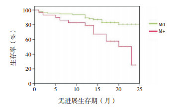
通过对M分期进行Kaplan-Meier分析:分期为M+者,中位PFS期为20(13~23)个月,20个月PFS率为50%±11%;分期为M0者20个月PFS率为81%±5%,与M+组相比,差异有统计学意义(P=0.001)。见图 1。

|
图 1 儿童MB M分期的生存分析曲线 |
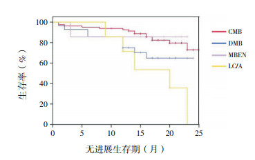
病理分型为CMB、DMB、MBEN、LC/A的20个月PFS率分别为80%±5%、65%±10%、86%±13%、36%±20%,不同病理分型之间PFS率差异有统计学意义(P=0.009),见图 2。

|
图 2 儿童MB不同病理分型的生存分析曲线 [CMB]经典型;[DMB]促纤维增生/结节型;[MBEN]广泛结节型;[LC/A]大细胞/间变型。 |
MB作为儿童最常见的神经系统肿瘤,虽然目前标危患儿5年生存率可达85%以上,高危患儿5年生存率约70%[10-11],但MB治疗后复发为影响MB预后的重要危险因素之一[12]。有研究提示,MB中位复发时间为23个月[13],故本文中重点研究与儿童MB两年内复发有关的危险因素。
Sabel等[14]对338例标危MB患儿随访10年发现,72例患儿出现复发,平均复发时间为32个月,中位复发时间为26个月。Huang等[15]研究显示MB的中位复发时间为17个月,且复发后中位生存时间为6个月,CMB与LC/A的5年PFS率分别为76%和34%,5年总体生存率分别为56%和76%,差异有统计学意义,提示LC/A预后较CMB差,且复发、转移率明显高于CMB。本研究复发率为24.4%,中位复发时间18个月,与以上研究结果相近。有些学者认为目前DMB与CMB预后哪个更好尚有争议,但小年龄组中,DMB的预后与MBEN相仿,均较CMB和LC/A要好[16-17],而本组病例中DMB与MBEN及CMB之间的差异无统计学意义,可能与DMB及MBEN病例数较少有关。
Rutkowski等[18]对5个中心270例5岁以下MB患儿进行回顾性分析发现,分期为M+者,其复发风险为M0者的2.28倍,与本研究结果相近,该研究略低的原因可能与其研究对象为5岁以下患儿有关。且该研究发现,与CMB相比,LC/A为预后差的危险因素,与本研究结果相符。
目前研究显示,儿童MB中WNT约占10%,SHH约30%,Group3约20%,Group4约40%[19]。本组病例中Group3占比相对较少可能与行分子分型的例数较少有关。WNT型MB的预后相对其他分型要好,该型患儿5年生存率可达95%[7]。本组研究中WNT型并未表现出明显的低风险,Group3型也未表现出更高的风险,可能与随访时间较短及病例数较少有关。
Yu等[20]通过对40例MB患儿的回顾性分析发现,起病时症状数量、M分期及术后放疗与生存预后相关,起病时症状数≥2者,其死亡风险为起病时症状<2者的2.61倍,而分期为M+者,其死亡风险为M0者的20.76倍。M+组患儿的复发风险明显高于M0组,为复发的独立危险因素,与本研究结果一致,但其复发风险较本研究高,可能与其随访时间较长有关,而应用Cox回归模型分析疾病预后危险因素时,分子分型对预后无影响,与本研究结果一致。
本组病例中,分期为M+者,20个月PFS率为50%±11%,明显低于M0。Verlooy等[21]研究发现,分期为M+者,5年PFS率为43.1%。Kortmann等[22]研究发现分期为M2~3者,3年PFS率为30%,结果均提示分期为M+预后不佳,与本文结果一致,但其PFS更低,可能与本组病例随访时间较短有关。Du等[23]研究表明,CMB 5年PFS率为29.3%±7.1%,DMB 5年PFS率为40.0%±15.5%,LC/A 3年PFS率为11.1%±0.5%,不同病理类型之间差异有统计学意义。与本组研究结论一致,但由于本组病例随访时间尚短,故无法将两组病例之间的PFS率进行比较。复发后患儿预后极差,3年存活率不足20%[14]。Perez-Somarriba等[24]报道,某患儿在复发后口服依托泊苷[50 mg/(m2·d),服用21 d,每4周为1个治疗周期]维持治疗,复发25个月后仍可继续上学并像复发前维持正常生活,提示虽然目前对于复发患儿可采取免疫治疗、靶向治疗等新型治疗,但传统化疗药物仍可选择。
MB为儿童最常见的脑恶性肿瘤,恶性程度高,虽然目前在多种方式的综合治疗下,其5年生存率有了明显的提高,但是复发后疾病的预后并不理想,本研究着重对两年内复发的危险因素进行回顾性分析,在今后的治疗过程中,应加强对有以上高危因素患儿的监管,进一步减少复发,提高患儿的远期生存率。
| [1] |
Dolecek TA, Propp JM, Stroup NE, et al. CBTRUS statistical report:primary brain and central nervous system tumors diagnosed in the United States in 2005-2009[J]. Neuro Oncol, 2012, 14(Suppl 5): v1-v49. DOI:10.1093/neuonc/nos218 (  0) 0) |
| [2] |
陈立华, 刘运生, 袁贤瑞, 等. 儿童髓母细胞瘤显微手术治疗[J]. 中国当代儿科杂志, 2002, 4(6): 478-481. DOI:10.3969/j.issn.1008-8830.2002.06.016 (  0) 0) |
| [3] |
Johnston DL, Keene D, Strother D, et al. Survival following tumor recurrence in children with medulloblastoma[J]. J Pediatr Hematol Oncol, 2018, 40(3): e159-e163. DOI:10.1097/MPH.0000000000001095 (  0) 0) |
| [4] |
Friedrich C, von Bueren AO, von Hoff K, et al. Treatment of adult nonmetastatic medulloblastoma patients according to the paediatric HIT 2000 protocol:a prospective observational multicentre study[J]. Eur J Cancer, 2013, 49(4): 893-903. DOI:10.1016/j.ejca.2012.10.006 (  0) 0) |
| [5] |
廖宇翔, 刘博, 李健, 等. 髓母细胞瘤复发相关因素分析及治疗方式探讨[J]. 国际神经病学神经外科学杂志, 2018, 45(3): 234-237. (  0) 0) |
| [6] |
Koschmann C, Bloom K, Upadhyaya S, et al. Survival after relapse of medulloblastoma[J]. J Pediatr Hematol Oncol, 2016, 38(4): 269-273. DOI:10.1097/MPH.0000000000000547 (  0) 0) |
| [7] |
Gajjar A, Chintagumpala M, Ashley D, et al. Risk-adapted craniospinal radiotherapy followed by high-dose chemotherapy and stem-cell rescue in children with newly diagnosed medulloblastoma (St Jude Medulloblastoma-96):long-term results from a prospective, multicentre trial[J]. Lancet Oncol, 2006, 7(10): 813-820. DOI:10.1016/S1470-2045(06)70867-1 (  0) 0) |
| [8] |
Northcott PA, Korshunov A, Witt H, et al. Medulloblastoma comprises four distinct molecular variants[J]. J Clin Oncol, 2011, 29(11): 1408-1414. DOI:10.1200/JCO.2009.27.4324 (  0) 0) |
| [9] |
Chang CH, Housepian EM, Herbert C Jr. An operative staging system and a megavoltage radiotherapeutic technic for cerebellar medulloblastomas[J]. Radiology, 1969, 93(6): 1351-1359. DOI:10.1148/93.6.1351 (  0) 0) |
| [10] |
Tarbell NJ, Friedman H, Polkinghorn WR, et al. High-risk medulloblastoma:a pediatric oncology group randomized trial of chemotherapy before or after radiation therapy (POG 9031)[J]. J Clin Oncol, 2013, 31(23): 2936-2941. DOI:10.1200/JCO.2012.43.9984 (  0) 0) |
| [11] |
Packer RJ, Gajjar A, Vezina G, et al. Phase Ⅲ study of craniospinal radiation therapy followed by adjuvant chemotherapy for newly diagnosed average-risk medulloblastoma[J]. J Clin Oncol, 2006, 24(25): 4202-4208. DOI:10.1200/JCO.2006.06.4980 (  0) 0) |
| [12] |
Pizer B, Donachie PH, Robinson K, et al. Treatment of recurrent central nervous system primitive neuroectodermal tumours in children and adolescents:results of a Children's Cancer and Leukaemia Group study[J]. Eur J Cancer, 2011, 47(9): 1389-1397. DOI:10.1016/j.ejca.2011.03.004 (  0) 0) |
| [13] |
Packer RJ, Zhou T, Holmes E, et al. Survival and secondary tumors in children with medulloblastoma receiving radiotherapy and adjuvant chemotherapy:results of Children's Oncology Group trial A9961[J]. Neuro Oncol, 2013, 15(1): 97-103. DOI:10.1093/neuonc/nos267 (  0) 0) |
| [14] |
Sabel M, Fleischhack G, Tippelt S, et al. Relapse patterns and outcome after relapse in standard risk medulloblastoma:a report from the HIT-SIOP-PNET4 study[J]. J Neurooncol, 2016, 129(3): 515-524. DOI:10.1007/s11060-016-2202-1 (  0) 0) |
| [15] |
Huang PI, Lin SC, Lee YY, et al. Large cell/anaplastic medulloblastoma is associated with poor prognosis-a retrospective analysis at a single institute[J]. Childs Nerv Syst, 2017, 33(8): 1285-1294. DOI:10.1007/s00381-017-3435-9 (  0) 0) |
| [16] |
Aref D, Croul S. Medulloblastoma:recurrence and metastasis[J]. CNS Oncol, 2013, 2(4): 377-385. DOI:10.2217/cns.13.30 (  0) 0) |
| [17] |
Siegfried A, Bertozzi AI, Bourdeaut F, et al. Clinical, pathological, and molecular data on desmoplastic/nodular medulloblastoma:case studies and a review of the literature[J]. Clin Neuropathol, 2016, 35(3): 106-113. (  0) 0) |
| [18] |
Rutkowski S, von Hoff K, Emser A, et al. Survival and prognostic factors of early childhood medulloblastoma:an international meta-analysis[J]. J Clin Oncol, 2010, 28(33): 4961-4968. DOI:10.1200/JCO.2010.30.2299 (  0) 0) |
| [19] |
Khatua S, Song A, Citla Sridhar D, et al. Childhood medulloblastoma:current therapies, emerging molecular landscape and newer therapeutic insights[J]. Curr Neuropharmacol, 2018, 16(7): 1045-1058. DOI:10.2174/1570159X15666171129111324 (  0) 0) |
| [20] |
Yu J, Zhao R, Shi W, et al. Risk factors for the prognosis of pediatric medulloblastoma:a retrospective analysis of 40 cases[J]. Clinics (Sao Paulo), 2017, 72(5): 294-304. DOI:10.6061/clinics/2017(05)07 (  0) 0) |
| [21] |
Verlooy J, Mosseri V, Bracard S, et al. Treatment of high risk medulloblastomas in children above the age of 3 years:a SFOP study[J]. Eur J Cancer, 2006, 42(17): 3004-3014. DOI:10.1016/j.ejca.2006.02.026 (  0) 0) |
| [22] |
Kortmann RD, Kühl J, Timmermann B, et al. Postoperative neoadjuvant chemotherapy before radiotherapy as compared to immediate radiotherapy followed by maintenance chemotherapy in the treatment of medulloblastoma in childhood:results of the German prospective randomized trial HIT'91[J]. Int J Radiat Oncol Biol Phys, 2000, 46(2): 269-279. DOI:10.1016/S0360-3016(99)00369-7 (  0) 0) |
| [23] |
Du S, Yang S, Zhao X, et al. Clinical characteristics and outcome of children with relapsed medulloblastoma:a retrospective study at a single center in China[J]. J Pediatr Hematol Oncol, 2018, 40(8): 598-604. DOI:10.1097/MPH.0000000000001241 (  0) 0) |
| [24] |
Perez-Somarriba M, Andión M, López-Pino MA, et al. Old drugs still work! Oral etoposide in a relapsed medulloblastoma[J]. Childs Nerv Syst, 2019, 35(5): 865-869. DOI:10.1007/s00381-019-04072-9 (  0) 0) |
 2019, Vol. 21


