 PDF(1158 KB)

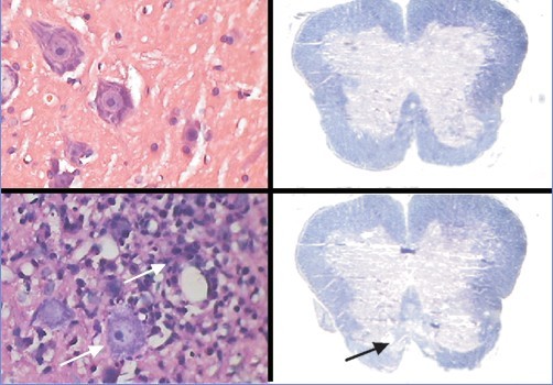
Features of pathological changes in the non-myelin sheath of rats with experimental autoimmune encephalomyelitis
ZHANG Jin-Feng, HUANG Rong,YANG Yu-Jia, XU Jun, JIN Shi-Jie
Chinese Journal of Contemporary Pediatrics ›› 2012, Vol. 14 ›› Issue (4) : 306-309.
PDF(1158 KB)
PDF(1158 KB)
Features of pathological changes in the non-myelin sheath of rats with experimental autoimmune encephalomyelitis
Experimental autoimmune encephalomyelitis / Myelin / Axonal damage / Apoptosis / Rats


