Diagnostic value of ultrasonographic examination for hepatic steatosis in obese children

ZHANG Hong-Xi, YANG Hui-Ping, LAI Can, HE Jing, YE Jing-Jing, FU Jun-Fen

Chinese Journal of Contemporary Pediatrics ›› 2014, Vol. 16 ›› Issue (9) : 873-877.

PDF(1272 KB)
PDF(1272 KB)
Chinese Journal of Contemporary Pediatrics ›› 2014, Vol. 16 ›› Issue (9) : 873-877. DOI: 10.7499/j.issn.1008-8830.2014.09.001
CLINICAL RESEARCH

Diagnostic value of ultrasonographic examination for hepatic steatosis in obese children

  • ZHANG Hong-Xi1, YANG Hui-Ping2, LAI Can1, HE Jing1, YE Jing-Jing1, FU Jun-Fen1
Author information +
History +

Abstract

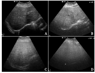
Objective To evaluate the sensitivity and specificity of hepatic ultrasonography(US) for the diagnosis of hepatic steatosis in obese children, using 1H magnetic resonance spectroscopy(1H MRS) as the reference standard.Methods A total of 162 obese children with age of 10.5±2.2 years and BMI of 28±4 were enrolled in this study. They accepted hepatic US and 1H MRS examinations. The sensitivity, specificity, positive predictive value(PPV) and negative predictive value(NPV) of US were calculated for the overall presence of hepatic steatosis by comparison with 1H MRS results. Results Using quantitative criteria of liver fat content(LFC) >5% determined by 1H MRS, 95 children(58.6%) were diagnosed as having hepatic steatosis. The sensitivity and specificity of US in diagnosing steatosis were 91.6%(87/95) and 50.7%(34/67) respectively, with PPV of 72.5%(87/120), and NPV of 81.0%(34/42). Considerable overlap in LFC measured by 1H MRS was observed between different grades from US findings: absent(LFC interquartile range:1.3%-3.9%), mild(2.4%-10.7%), moderate(7.1%-20.2%) and severe(7.6%-28.8%) steatosis. Conclusion The US can yield a high sensitivity and low specificity in the diagnosis of hepatic steatosis in obese children, suggesting it can be used as a screening tool for hepatic steatosis. To improve diagnostics, 1H MRS is needed to determine LFC.

Key words

Ultrasonography / 1H magnetic resonance spectroscopy / Obesity / Hepatic steatosis / Child

Cite this article

Download Citations
ZHANG Hong-Xi, YANG Hui-Ping, LAI Can, HE Jing, YE Jing-Jing, FU Jun-Fen. Diagnostic value of ultrasonographic examination for hepatic steatosis in obese children[J]. Chinese Journal of Contemporary Pediatrics. 2014, 16(9): 873-877 https://doi.org/10.7499/j.issn.1008-8830.2014.09.001

References

[1] Schwimmer JB, Deutsch R, Kahen T, et al.Prevalence of fatty liver in children and adolescents [J].Pediatrics, 2006, 118(4):1388-1393.
[2] Pacifico L, Nobili V, Anania C, et al.Pediatric nonalcoholic fatty liver disease, metabolic syndrome and cardiovascular risk[J].World J Gastroenterol, 2011, 17(26): 3082-3091.
[3] Pais R, Pascale A, Fedchuck L, et al.Progression from isolated steatosis to steatohepatitis and fibrosis in nonalcoholic fatty liver disease [J].Clin Res Hepatol Gastroenterol, 2011, 35(1): 23-28.
[4] Strauss S, Gavish E, Gottlieb P, et al.Interobserver and intraobserver variability in the sonographic assessment of fatty liver [J].AJR Am J Roentgenol, 2007, 189(6): W320-W323.
[5] Mehta SR, Thomas EL, Patel N, et al.Proton magnetic resonance spectroscopy and ultrasound for hepatic fat quantification [J].Hepatol Res, 2010, 40(4): 399-406.
[6] Longo R, Pollesello P, Ricci C, et al.Proton MR spectroscopy in quantitative in vivo determination of fat content in human liver steatosis [J].J Magn Reson Imaging, 1995, 5(3): 281-285.
[7] Roldan-Valadez E, Favila R, Martinez-Lopez M, et al.In vivo 3T spectroscopic quantification of liver fat content in nonalcoholic fatty liver disease: Correlation with biochemical method and morphometry [J].J Hepatol, 2010, 53(4): 732-737.
[8] van Werven JR, Hoogduin JM, Nederveen AJ, et al.Reproducibility of 3.0 Tesla magnetic resonance spectroscopy for measuring hepatic fat content [J].J Magn Reson Imaging,2009, 30(2): 444-448.
[9] Mancini M, Prinster A, Annuzzi G, et al.Sonographic hepatic-renal ratio as indicator of hepatic steatosis: comparison with(1)H magnetic resonance spectroscopy [J].Metabolism, 2009,58(12): 1724-1730.
[10] Perseghin G, Bonfanti R, Magni S, et al.Insulin resistance and whole body energy homeostasis in obese adolescents with fatty liver disease [J].Am J Physiol Endocrinol Metab, 2006, 291(4):E697-E703.
[11] 李辉, 季成叶, 宗心南, 等.中国 0~18 岁儿童、青少年体块指数的生长曲线 [J].中华儿科杂志, 2009, 47(7):493-498.
[12] 中华医学会肝脏病学分会脂肪肝和非酒精性肝病学组.非酒精性脂肪肝病诊疗指南 [J].中华肝脏病杂志, 2006, 14(3):161-163.
[13] Graif M, Yanuka M, Baraz M, et al.Quantitative estimation of attenuation in ultrasound video images: correlation with histology in diffuse liver disease [J].Invest Radiol, 2000, 35(5):319-324.
[14]张洪锡, 傅君芬, 黄轲, 等.非酒精性脂肪肝病儿童、青少年的肝内脂肪定量评价 [J].中国当代儿科杂志, 2012, 14(8):598-603.
[15] Mottin CC, Moretto M, Padoin AV, et al.The role of ultrasound in the diagnosis of hepatic steatosis in morbidly obese patients[J].Obes Surg, 2004, 14(5): 635-637.
[16] de Moura Almeida A, Cotrim HP, Barbosa DB, et al.Fatty liver disease in severe obese patients: diagnostic value of abdominal ultrasound [J].World J Gastroenterol, 2008, 14(9): 1415-1418.
PDF(1272 KB)

Accesses

Citation

Detail

Sections
Recommended

/