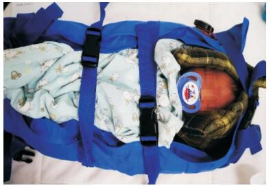
 PDF(1765 KB)


真空固定器联合喂奶方式在新生儿头颅磁共振成像中的应用——前瞻性随机对照研究
沈晓霞, 刘婷婷, 高福生, 吴丹, 杜立中, 马晓路
中国当代儿科杂志 ›› 2020, Vol. 22 ›› Issue (5) : 435-440.
 PDF(1765 KB)
 PDF(1765 KB)
真空固定器联合喂奶方式在新生儿头颅磁共振成像中的应用——前瞻性随机对照研究
Application of vacuum stretcher combined with feeding in cranial magnetic resonance imaging examination for neonates: a prospective randomized controlled study
目的 评价真空固定器联合喂奶方式在新生儿头颅磁共振成像(MRI)中的有效性和安全性。方法 前瞻性选择2019年9~11月浙江大学医学院附属儿童医院新生儿科需要进行头颅MRI检查的胎龄 > 34周且生命体征平稳、不需要吸氧的高胆红素血症新生儿为研究对象,随机分为真空固定器联合喂奶组和常规镇静组,所有患儿均在头颅MRI检查前后进行生命体征监测,记录MRI完成情况。结果 共纳入80例新生儿,真空固定器联合喂奶组40例,常规镇静组40例。真空固定器联合喂奶组头颅MRI检查成功率高于常规镇静组(92% vs 75%,P < 0.05)。扫描成功的患儿中,真空固定器联合喂奶组检查完成后的最快心率低于常规镇静组(P < 0.05);两组患儿在头颅MRI前后经皮血氧饱和度、呼吸频率、体温等差异无统计学意义(P > 0.05)。所有患儿均未发生呼吸暂停、急性过敏反应、恶性高热等并发症。结论 真空固定器联合喂奶方式在不增加风险的前提下提高了头颅MRI检查的成功率,有效减少镇静剂的使用。
Objective To study the effect and safety of vacuum stretcher combined with feeding in cranial magnetic resonance imaging (MRI) examination for neonates. Methods A prospective study was performed for the neonates with hyperbilirubinemia, with a gestational age of > 34 weeks and stable vital signs, who needed cranial MRI examination and did not need oxygen inhalation hospitalized in the Department of Neonatology, Children's Hospital of Zhejiang University School of Medicine, from September to November, 2019. The neonates were randomly divided into a vacuum stretcher combined with feeding group and a conventional sedation group. Vital signs were monitored before, during, and after MRI examination. The success rate of MRI procedure was recorded. Results A total of 80 neonates were enrolled in the study, with 40 neonates in the vacuum stretcher combined with feeding group and 40 in the conventional sedation group. The vacuum stretcher combined with feeding group had a significantly higher success rate of MRI procedure than the conventional sedation group (P < 0.05). As for the neonates who underwent successful MRI examination, the fastest heart rate after examination in the vacuum stretcher combined with feeding group was significantly lower than that in the conventional sedation group (P < 0.05), while there were no significant differences between the two groups in transcutaneous oxygen saturation, respiratory rate, and body temperature before and after MRI examination (P > 0.05). No complications, such as apnea, acute allergic reactions, and malignant fever, were observed. Conclusions Vacuum stretcher combined with feeding can improve the success rate of MRI procedure and reduce the use of sedatives, and meanwhile, it does not increase related risks.

磁共振成像 / 镇静剂 / 真空固定器 / 喂奶 / 新生儿
Magnetic resonance imaging / Sedative / Vacuum stretcher / Feeding / Neonate
[1] Jaimes C, Gee MS. Strategies to minimize sedation in pediatric body magnetic resonance imaging[J]. Pediatr Radiol, 2016, 46(6):916-927.
[2] Kang R, Shin YH, Gil NS, et al. A comparison of the use of propofol alone and propofol with midazolam for pediatric magnetic resonance imaging sedation-a retrospective cohort study[J]. BMC Anesthesiol, 2017, 17(1):138.
[3] Barton K, Nickerson JP, Higgins T, et al. Pediatric anesthesia and neurotoxicity:what the radiologist needs to know[J]. Pediatr Radiol, 2018, 48(1):31-36.
[4] Bjur KA, Payne ET, Nemergut ME, et al. Anesthetic-related neurotoxicity and neuroimaging in children:a call for caonversation[J]. J Child Neurol, 2017, 32(6):594-602.
[5] Parad RB. Non-sedation of the neonate for radiologic procedures[J]. Pediatr Radiol, 2018, 48(4):524-530.
[6] Shariat M, Mertens L, Seed M, et al. Utility of feed-and-sleep cardiovascular magnetic resonance in young infants with complex cardiovascular disease[J]. Pediatr Cardiol, 2015, 36(4):809-812.
[7] Shariat M, Mertens L, Seed M, et al. Utility of feed-and-sleep cardiovascular magnetic resonance in young infants with complex cardiovascular disease[J]. Pediatr Cardiol, 2015, 36(4):809-812.
[8] Ahmad R, Hu HH, Krishnamurthy R, et al. Reducing sedation for pediatric body MRI using accelerated and abbreviated imaging protocols[J]. Pediatr Radiol, 2018, 48(1):37-49.
[9] Kannikeswaran N, Mahajan PV, Sethuraman U, et al. Sedation medication received and adverse events related to sedation for brain MRI in children with and without developmental disabilities[J]. Paediatr Anaesth, 2009, 19(3):250-256.
[10] Jaimes C, Murcia DJ, Miguel K, et al. Identification of quality improvement areas in pediatric MRI from analysis of patient safety reports[J]. Pediatr Radiol, 2018, 48(1):66-73.
[11] Khawaja AA, Tumin D, Beltran RJ, et al. Incidence and causes of adverse events in diagnostic radiological studies requiring anesthesia in the Wake-Up Safe registry[J]. J Patient Saf, 2018. DOI:10.1097/PTS.0000000000000469. Epub ahead of print.
[12] Antonov NK, Ruzal-Shapiro CB, Morel KD, et al. Feed and wrap MRI technique in infants[J]. Clin Pediatr (Phila), 2017, 56(12):1095-1103.
[13] Ibrahim T, Few K, Greenwood R, et al. ‘Feed and wrap’ or sedate and immobilise for neonatal brain MRI?[J]. Arch Dis Child Fetal Neonatal Ed, 2015, 100(5):F465-F466.
[14] Kini K, Kini PG. Sleep deprivation for radiological procedures in children[J]. Pediatr Radiol, 2009, 39(11):1255.
[15] Nelson AM. Risks and benefits of swaddling healthy infants:an integrative review[J]. MCN Am J Matern Child Nurs, 2017, 42(4):216-225.
[16] van Sleuwen BE, Engelberts AC, Boere-Boonekamp MM, et al. Swaddling:a systematic review[J]. Pediatrics, 2007, 120(4):e1097-e1106.
[17] Edwards AD, Arthurs OJ. Paediatric MRI under sedation:is it necessary? What is the evidence for the alternatives?[J]. Pediatr Radiol, 2011, 41(11):1353-1364.
[18] Mahan ST, Kasser JR. Does swaddling influence developmental dysplasia of the hip?[J]. Pediatrics, 2008, 121(1):177-178.
[19] Linder JMB. Safety considerations in immobilizing pediatric clients for radiographic procedures[J]. J Radiol Nurs, 2017, 36(1):55-58.