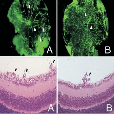
摘要 目的:探讨特异性抑制缺氧诱导因子-1α(HIF-1α)的表达对早产儿视网膜病变(retinopathy of prematurity,ROP)小鼠模型视网膜新生血管的抑制作用。方法:新生C57BL/6J小鼠48只,随机分为实验组和对照组(每组24只),采用Smith方法制备ROP模型。生后12 d 时实验组玻璃体腔注射HIF-1α特异性小片段干扰RNA 表达载体pSUPERH1-siHIF-1α,同时对照组玻璃体注射空载体。Western blot检测两组小鼠视网膜HIF-1α和血管内皮生长因子(VEGF)的表达,FITC-Dextran荧光照影视网膜铺片和组织切片观察两组小鼠视网膜新生血管和新生血管内皮细胞核数目的差异。结果:实验组小鼠HIF-1α和VEGF的表达较对照组明显下降(P<0.01);实验组突破视网膜内界膜血管内皮细胞核数较对照组明显减少(P<0.01)。结论:特异性抑制HIF-1α能有效抑制ROP视网膜新生血管的形成。
Abstract:OBJECTIVE: To study the inhibition effect of HIF-1α specific siRNA expression vector pSUPERH1-siHIF-1α on retinal neovascularization in a mouse model of retinopathy of prematurity (ROP). METHODS: The mouse model of ROP was prepared by the method Smith described. Forty-eight ROP mice were randomly divided into two groups: an experimental group that was intravitreously injected with pSUPERH1-siHIF-1α and a control group that was injected with pSUPER retro vector. The levels of HIF-1α and vascular endothelia growth factor (VEGF) in the retina were examined by Western blot. The retinal neovascularization was evaluated by angiography using FITC Dextran and quantitated histologically. RESULTS: The levels of HIF-1α and VEGF in the retina in the experimental group were reduced 90% and 65% respectively compared with those in the control group. Meanwhile, the number of retinal neovascular endothelial nucleus outbreaking the inner limit membrane in the experimental group was significantly reduced compared with that in the control group. CONCLUSIONS: The development of retinal neovascularization of ROP can be markedly inhibited by RNA interference targeting HIF-1α.
XU Hui-Zhuo,LIU Shuang-Zhen,XIONG Si-Qi et al. HIF-1α siRNA reduces retinal neovascularization in a mouse model of retinopathy of prematurity[J]. CJCP, 2011, 13(8): 680-683.
[1]The Committee for the Classification of Retinopathy of Prematurity. An international classification of retinopathy of prematurity[J]. Arch Ophthalmol, 1984,102(8): 1130-1134.
[3]Simó R, Carrasco E, García-Ramírez M, Hernández C. Angiogenic and antiangiogenic factors in proliferative diabetic retinopathy[J]. Curr Diabetes Rev, 2006, 2(1):71-98.
[4]Robinson GS, Pierce EA, Rook SL, Foley E, Webb R, Smith LE. Oligodeoxynucleotides inhibit retinal neovascularization in a murine model of proliferative retinopathy[J]. Proc Natl Acad Sci U S A, 1996, 93(10): 4851-4856.
[5]Bainbridge JW, Mistry A, De Alwis M, Paleolog E, Baker A, Thrasher AJ, et al. Inhibition of retinal neovascularisation by gene transfer of soluble VEGF receptor sFlt1[J]. Gene Ther, 2002, 9(5): 320-326.
[6]Rosmorduc O, Housset C. Hypoxia: a link between fibrogenesis, angiogenesis, and carcinogenesis in liver disease[J]. Semin Liver Dis, 2010, 30(3): 258-270.
[7]Westra J, Molema G, Kallenberg CG. Hypoxia-inducible factor-1 as regulator of angiogenesis in rheumatoid arthritis-therapeutic implications[J]. Curr Med Chem, 2010, 17(3): 254-263.
[10]Smith LE, Wesolowski E, McLellan A, Kostyk SK, D’Amato R, Sullivan R, et al. Oxygen-induced retinopathy in the mouse[J]. Invest Ophthalmol Vis Sci, 1994, 35(1): 101-111.
[11]Ozaki H, Yu AY, Della N, Ozaki K, Luna JD, Yamada H, et al. Hypoxia inducible factor-1alpha is increased in ischemic retina: temporal and spatial correlation with VEGF expression[J]. Invest Ophthalmol Vis Sci, 1999, 40(1): 182-189.
[12]Masuda I, Matsuo T, Yasuda T, Matsuo N. Gene transfer with the intraocular tissues by different routes of administration[J]. Invest Ophthalmol Vis Sci, 1996, 345(37): 1914-1920.
[13]Lipardi C, Wei Q, Paterson BM. RNAi as random degradative PCR: siRNA primers convert mRNA into dsRNAs that are degraded to generate new siRNAs[J]. Cell, 2001, 107(3): 297-307.
[14]Takahashi R, Tanaka S, Hiyama T, Ito M, Kitadai Y, Sumii M, et al. Hypoxia-inducible factor-1alpha expression and angiogenesis in gastrointestinal stromal tumor of the stomach[J]. Oncol Rep, 2003, 10(4): 797-802.
[15]Frank RN. Diabetic retinopathy[J]. N Engl J Med, 2004, 350(1):48-58.