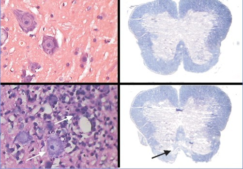
目的:探讨实验性自身免疫性脑脊髓炎(EAE)大鼠非髓鞘组织病理变化的特征及临床意义。方法:48只Wistar大鼠随机分为对照组(CON,24只)和EAE组(24只),比较两组大鼠行为学;用苏木精-伊红染色、LOYEZ髓鞘染色、免疫组化及TUNEL方法观察EAE大鼠脊髓的炎症反应、脱髓鞘改变、神经丝蛋白和神经元的损伤。结果:行为学及组织学证实EAE大鼠模型复制成功。同时发现EAE组大鼠免疫诱导7 d后神经丝蛋白密度开始减低,排列紊乱,可见轴索断裂及轴索脱失,14 d达高峰,免疫诱导7 d、14 d及21 d神经丝蛋白密度均明显低于对照组,差异有统计学意义(P< 0.01);免疫诱导7 d、14 d及21 d神经元的凋亡指数与对照组相比明显升高,差异有统计学意义(P< 0.01)。结论:EAE大鼠模型除了炎性脱髓鞘病变外,在疾病早期开始出现明显轴索损伤及神经元的凋亡,与神经功能障碍有关。
Abstract
OBJECTIVE: To study the pathological changes in the non-myelin sheath by observing histological damages to the neurofilament protein and apoptosis of neurons in rats with experimental autoimmune encephalomyelitis (EAE). METHODS: Forty-eight Wistar rats were randomly divided into two groups: control and EAE (24 rats in each group). Behavioral changes were observed. Inflammation reactions and demyelination were observed by hematoxylin eosin staining and LOYEZ staining.The level of neurofilament was detected by immunohistochemistry. Apoptosis of the neuron in the spinal cord was detected by TUNEL. RESULTS: Behavioral and histological results confirmed that the model of EAE rats was prepared successfully. In the EAE group, typical morphological features of axonal damage (sparsed axonal density, axonal distortion, axonal transection and even axonal disappearance) were found from the seventh day after immunization and the morphological changes were the most obvious on the fourteenth day. Neurofilament density in the EAE group was significantly lower than in the control group (P<0.01) at 7, 14 and 21 days after immunization. The neuronal apoptosis index in the EAE group at 7, 14 and 21 days after immunization was significantly higher than in the control group (P<0.01). CONCLUSIONS: In addition to inflammatory demyelination, axonal damage and neuronal apoptosis can be observed in the early stage of EAE. Pathological changes may be associated with neurological dysfunction.
关键词
实验性变态反应性脑脊髓炎 /
髓鞘 /
轴索损伤 /
凋亡 /
大鼠
Key words
Experimental autoimmune encephalomyelitis /
Myelin /
Axonal damage /
Apoptosis /
Rats
{{custom_sec.title}}
{{custom_sec.title}}
{{custom_sec.content}}
参考文献
[1]Ercolini AM, Miller SD. Mechanisms of immunopathology in murine models of central nervous system demyelinating disease[J]. J Immunol, 2006, 176(6): 3293-3298.
[2]De Stefano N, Filippi M. MR spectroscopy in multiple sclerosis[J]. J Neuroimaging, 2007, 17(suppl 1): 31s-35s.
[3]Haghighi S, Andersen O, Odén A, Rosengren L. Cerebrospinal fluid markers in MS patient s and their healthy siblings[J]. Acta Neurol Scand, 2004, 109(2): 97-99.
[4]Lassmann H. Axonal and neuronal pathology in multiple sclerosis: what have we learnt from animal models[J]. Exp Neurol, 2010, 225(1): 2-8.
[5]Benson JM,Campbell KA,Guan Z, Gienapp IE, Stuckman SS, Forsthuber T, et al. T-cell activation and receptor downmodulation precede deletion induced by mucosally administered antigen[J]. J Clin Invest, 2000, 106(8): 1031-1038.
[6]Rovaris M, Gambini A, Gallo A, Falini A, Ghezzi A, Benedetti B, et al. Axonal injury in early multiple sclerosis is irreversible and independent of the shortterm disease evolution[J]. Neurology, 2005, 65(10): 1626-1630.
[7]Lee MA, Blamire AM, Pendlebury S, HO KH, Mills KR, Styles P, et al. Axonal injury or loss in the internal capsule and motor impairment in multiple sclerosis[J]. Arch Neurol, 2000, 57(1): 65-70.
[8]李宏增, 林宏, 宿长军, 李柱一, 孟民杰.髓鞘碱性蛋白反应性淋巴细胞对神经细胞的作用[J].中华神经外科疾病研究杂志, 2006, 5(3): 233-235.
[9]Linker RA, Sendtner M, Gold R. Mechanisms of axonal degeneration in EAE-lessons from CNTF and MHC I knockout mice[J]. J Neurol Sci, 2005, 233(1-2): 167-172.
[10]徐俊,黄榕,杨于嘉,金世杰,张金凤. 黄芩苷对自身免疫性脑脊髓炎大鼠细胞凋亡的影响[J]. 中国当代儿科杂志,2011,13(8):665-668.
[11]Wattjes MP, Harzheim M, Lutterbey GG, Klotz L, Schild HH, Tr-ber F. Axonal damage but no increased glial cell activity in the normal-appearing white matter of patients with clinically isolated syndromes suggestive of multiple sclerosis using high-field magnetic resonance spectroscopy[J]. AJNR Am J Neuroradiol, 2007, 28(8): 1517-1522.
[12]Niki'c I, Merkler D, Sorbara C, Brinkoetter M, Kreutzfeldt M, Bareyre FM, et al. A reversible form of axon damage in experimental autoimmune encephalomyelitis and multiple sclerosis[J]. Nat Med, 2011, 17(4): 495-499.
 PDF(1158 KB)