新生大鼠缺氧缺血脑损伤时脑组织STAT3信号通路的作用

邓睿, 赵凤艳, 张莉, 李德渊, 母得志

中国当代儿科杂志 ›› 2016, Vol. 18 ›› Issue (1) : 78-84.

PDF(3162 KB)
HTML
PDF(3162 KB)
HTML
中国当代儿科杂志 ›› 2016, Vol. 18 ›› Issue (1) : 78-84. DOI: 10.7499/j.issn.1008-8830.2016.01.016
论著·实验研究

新生大鼠缺氧缺血脑损伤时脑组织STAT3信号通路的作用

  • 邓睿1,2, 赵凤艳1, 张莉1, 李德渊1, 母得志1
作者信息 +

Role of STAT3 signaling pathway in hypoxic-ischemic brain damage of neonatal rats

  • DENG Rui1,2, ZHAO Feng-Yan1, ZHANG Li1, LI De-Yuan1, MU De-Zhi1
Author information +
文章历史 +

摘要

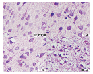
目的 研究信号转导和转录激活因子3(STAT3)信号通路在新生大鼠缺氧缺血脑损伤中的作用和机制。方法 80 只7 日龄Sprague-Dawley 大鼠随机分为缺氧缺血(HI) 组(n=40)和假手术组(n=40)。HI 组大鼠行右侧颈总动脉结扎随后缺氧(8% O2)处理2.5 h,假手术组仅分离右侧颈总动脉但不予结扎和缺氧处理。两组分别在HI 后4、6、8、12 和24 h 处死大鼠并收集脑组织。采用免疫组化和Westernblot 法检测STAT3、磷酸化STAT3 和血管内皮生长因子(VEGF)蛋白表达,TUNEL 染色法检测细胞凋亡。结果 HI 组和假手术组间STAT3 蛋白表达在各时间点差异均无统计学意义(P>0.05);除24 h 外,其余时间点HI 组的磷酸化STAT3 蛋白表达均明显高于假手术组,6 h 达高峰(P<0.01)。各时间点HI 组VEGF 蛋白表达均明显高于假手术组,8 h 达高峰(P<0.05)。HI 组各时间点凋亡细胞数均高于假手术组,且随时间延长凋亡细胞数逐渐增加(P<0.01)。结论 新生大鼠HI 后STAT3 可能被磷酸化激活从而诱导VEGF 表达;STAT3通路激活可能参与了神经细胞的凋亡调节,推测该通路活化与神经细胞的凋亡抑制相关。

Abstract

Objective To study the role and mechanisms of STAT3 signaling pathway in hypoxic-ischemic brain damage (HIBD) of neonatal rats. Methods Eighty 7-day-old Sprague-Dawley rats were randomly divided into two groups: HI and sham-operated (n=40 each). The rats in the HI group were subjected to right carotid artery ligation and subsequent hypoxia exposure (8% O2) for 2.5 hours, and the rats in the sham-operated group underwent the right carotid artery dissection without subsequent ligation or hypoxia treatment. Brain tissue samples were collected at 4, 6, 8, 12 and 24 hours after operation and hypoxic exposure. Immunohistochemistry and Western blot were used to detect the expression of STAT3, phosphorylated STAT3 (p-STAT3) and vascular endothelial growth factor (VEGF) proteins. TUNEL staining was used to detect apoptotic cells. Results No significant difference in STAT3 expression was observed at all time points between the HI and sham-operated groups (P>0.05). Compared with the sham-operated group, the expression of p-STAT3 protein in the HI group was significantly upregulated at 4, 6, 8, 12 hours after operation and hypoxic exposure, and peaked at 6 hours (P<0.01). The VEGF expression in the HI group was higher than that in the sham-operated group at all time points, which peaked at 8 hours (P<0.05). TUNEL staining showed that the apoptotic cells increased significantly in a time-dependent manner compared with the sham-operated group (P<0.01). Conclusions HI may lead to phosphorylation of STAT3 which probably induces the VEGF expression in the brain of neonatal rats. The activated STAT3 signaling pathway may be involved in the apoptosis regulation of nerve cells, and related to apoptosis inhibition of nerve cells.
关键词

信号转导和转录激活因子3 / 血管内皮生长因子 / 凋亡 / 缺氧缺血性脑损伤 / 新生大鼠

Key words

Signal transducer and activator of transcription 3 / Vascular endothelial growth factor / Apoptosis / Hypoxic-ischemic brain damage / Neonatal rats

引用本文

导出引用
邓睿, 赵凤艳, 张莉, 李德渊, 母得志. 新生大鼠缺氧缺血脑损伤时脑组织STAT3信号通路的作用[J]. 中国当代儿科杂志. 2016, 18(1): 78-84 https://doi.org/10.7499/j.issn.1008-8830.2016.01.016
DENG Rui, ZHAO Feng-Yan, ZHANG Li, LI De-Yuan, MU De-Zhi. Role of STAT3 signaling pathway in hypoxic-ischemic brain damage of neonatal rats[J]. Chinese Journal of Contemporary Pediatrics. 2016, 18(1): 78-84 https://doi.org/10.7499/j.issn.1008-8830.2016.01.016

参考文献

[1] 谢欣梅, 庞晓斌, 赵艳, 等. 消栓通络有效成分组对氧糖剥夺 损伤原代培养神经元的保护作用[J]. 药学学报, 2014, 49(8): 1130-1135.
[2] Zhou H, Zhang Z, Wei H, et al. Activation of STAT3 is involved in neuroprotection by electroacupuncture pretreatment via cannabinoid CB1 receptors in rats[J]. Brain Res, 2013, 1529: 154-164.
[3] Lin Y, Cai B, Xue XH, et al. TAT-mediated delivery of neuroglobin attenuates apoptosis induced by oxygen-glucose deprivation via the Jak2/Stat3 pathway in vitro[J]. Neurol Res, 2015, 37(6): 531-538.
[4] Liu R, Xu N, Yi W, et al. Electroacupuncture effects on cortical neurons, as well as Janus kinase 2-signal transducer and activator of transcription 3 signal transduction pathway, in a rat model of cerebral ischemia[J]. Neural Regen Res, 2012, 7(6): 457-462.
[5] Meares GP, Liu Y, Rajbhandari R, et al. PERK-dependent activation of JAK1 and STAT3 contributes to endoplasmic reticulum stress-induced inflammation[J]. Mol Cell Biol, 2014, 34(20): 3911-3925.
[6] Cai B, Cai JP, Luo YL, et al. The specific roles of JAK/STAT signaling pathway in sepsis[J]. Inflammation, 2015, 38(4): 1599-1608.
[7] Hong S, Song MR. STAT3 but not STAT1 is required for astrocyte differentiation[J]. PLoS One, 2014, 9(1): e86851.
[8] Song S, Park JT, Na JY, et al. Early expressions of hypoxiainducible factor 1alpha and vascular endothelial growth factor increase the neuronal plasticity of activated endogenous neural stem cells after focal cerebral ischemia[J]. Neural Regen Res, 2014, 9(9): 912-918.
[9] Gao L, Zhou S, Cai H, et al. VEGF levels in CSF and serum in mild ALS patients[J]. J Neurol Sci, 2014, 346(1-2): 216-220.
[10] Li L, Qu Y, Mao M, et al. The involvement of phosphoinositid 3-kinase/Akt pathway in the activation of hypoxia-inducible factor-1alpha in the developing rat brain after hypoxiaischemia[ J]. Brain Res, 2008, 1197: 152-158.
[11] Rice JE 3rd, Vannucci RC, Brierley JB. The influence of immaturity on hypoxic-ischemic brain damage in the rat[J]. Ann Neurol, 1981, 9(2): 131-141.
[12] Villarino AV, Kanno Y, Ferdinand JR, et al. Mechanisms of Jak/ STAT signaling in immunity and disease[J]. J Immunol, 2015, 194(1): 21-27.
[13] Yamashita T, Sawamoto K, Suzuki S, et al. Blockade of interleukin-6 signaling aggravates ischemic cerebral damage in mice: possible involvement of STAT3 activation in the protection of neurons[J]. J Neurochem, 2005, 94(2): 459-468.
[14] Suzuki S, Tanska K, Nogawa S, et al. Phosphorylation of signal transducer and activator of transcription-3 (STAT3) after focal cerebral ischemia in rats[J]. Exp Neurol, 2001, 170(1): 63-71.
[15] Kamran MZ, Patil P, Gude RP. Role of STAT3 in cancer metastasis and translational advances[J]. Biomed Res Int, 2013, 2013: Article ID 421821, 15 pages.
[16] Rosenstein JM, Krum JM, Ruhrberg C. VEGF in the nervous system[J]. Organogenesis, 2010, 6(2):107-114.
[17] Zhou TF, Yu JG. Recombinant human erythropoietin attenuates neuronal apoptosis and cognitive defects via JAK2/STAT3 signaling in experimental endotoxemia[J]. J Surg Res, 2013, 183(1): 304-312.
[18] 李丽华, 屈艺, 张莉, 等. HIF-lα 表达在新生大鼠缺氧/ 缺 氧缺血性脑损伤神经元凋亡中的作用[J]. 四川大学学报( 医 学版), 2008, 39(6): 912-915.

基金

国家教育部博士点基金(20120181120049);国家自然科学基金青年科学基金(81300526;81200462)。


PDF(3162 KB)
HTML

Accesses

Citation

Detail

段落导航
相关文章

/