 PDF(1453 KB)


骨髓间充质干细胞移植对氧诱导视网膜病变新生大鼠视网膜新生血管的影响
牟青杰, 赵月华, 程丹丹, 王海宇, 陈岚芬, 赵岩松, 王晓莉
中国当代儿科杂志 ›› 2017, Vol. 19 ›› Issue (11) : 1202-1207.
 PDF(1453 KB)
 PDF(1453 KB)
骨髓间充质干细胞移植对氧诱导视网膜病变新生大鼠视网膜新生血管的影响
Effects of bone marrow mesenchymal stem cell transplantation on retinal neovascularization in neonatal rats with oxygen-induced retinopathy
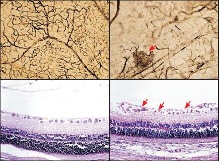
目的 探讨大鼠骨髓间充质干细胞(BMSC)移植对氧诱导视网膜病变(OIR)新生大鼠视网膜新生血管的影响,并观察视网膜缺氧诱导因子-1α(HIF-1α)、血管内皮生长因子(VEGF)表达的变化,为BMSC临床治疗OIR提供可靠的理论依据。方法 将72只7日龄Sprague-Dawley新生大鼠随机分为正常对照组、模型组(OIR组)与BMSC移植组(BMSC组)。体外培养BMSC,采用氧诱导法制成OIR模型,持续高氧5 d后,BMSC组行玻璃体内注射大鼠BMSC。移植后7 d,采用视网膜铺片法、苏木精-伊红染色(HE)法检测BMSC移植对OIR新生大鼠视网膜新生血管的影响;免疫组化法结合Western blot法观察BMSC对OIR新生大鼠视网膜HIF-1α及VEGF蛋白表达的影响。结果 视网膜铺片结果示正常对照组视网膜血管排列整齐,而OIR组血管排列紊乱,可见大片无灌注区,BMSC组仅可见大血管稍迂曲,血管排列较规整,无灌注区明显减少;HE染色发现OIR组视网膜内界膜可见大量新生血管及视网膜前新生血管细胞(Pre-RNC),BMSC组视网膜内界膜新生血管明显减少,且pre-RNC数显著少于OIR组(P < 0.01);免疫组织化学结果示OIR组HIF-1α阳性与VEGF阳性细胞数显著多于正常对照组,而BMSC组HIF-1α阳性与VEGF阳性细胞数均显著少于OIR组(P < 0.05)。Western blot结果示OIR组HIF-1α与VEGF蛋白表达显著高于正常对照组,而BMSC组HIF-1α与VEGF蛋白表达显著低于OIR组(P < 0.01)。结论 BMSC玻璃体内移植可减轻OIR新生大鼠视网膜新生血管的形成,其机制可能与其抑制HIF-1α及VEGF蛋白的表达有关。
Objective To explore the effects of rat bone mesenchymal stem cell (BMSC) transplantation on retinal neovascularization, and to observe the changes of hypoxia-inducible factor-1 alpha (HIF-1α) and vascular endothelial growth factors (VEGF) in rats with oxygen-induced retinopathy (OIR). Methods Seventy-two seven-day-old Sprague-Dawley rats were randomly divided into three groups:normal control (CON), model (OIR) and BMSC transplantation. In the BMSC transplantation group, BMSCs were transplanted 5 days after oxygen conditioning. The phosphate buffered saline of the same volume was injected in the CON and OIR groups. The OIR model was prerpared according to the classic hyperoxygen method. At seven days after transplantation, retinal neovascularization was examined by retinal flat-mount staining and hematoxylin eosin (HE) staining. The expression of HIF-1α and VEGF proteins was examined by immunohistochemistry staining and Western blot analysis. Results The retinal flat-mount staining results showed that the vessels were well organized in the CON group, but the vessels were irregularly organized, and lots of nonperfusion areas were observed in the OIR group. The large vessels were a bit circuitous, the retinal vessels were relatively organized, and less nonperfusion areas were noted in the BMSC transplantation group. The HE staining results showed that many neovessels and preretinal neovascular (pre-RNC) cells were observed on the internal limiting membrane in the OIR group. There were less pre-RNC cells in the BMSC transplantation group compared with the OIR group (P < 0.01). The immunohistochemistry analysis showed that more HIF-1α+ and VEGF+ cells were observed in the OIR group compared with the CON group, and less HIF-1α+ and VEGF+ cells were observed in the BMSC transplantation group compared with OIR group (P < 0.05). The Western blot analysis showed the expression of HIF-1α and VEGF proteins in the OIR group was significantly higher than that in the CON group. The expression of HIF-1α and VEGF proteins in the BMSC transplantation group was lower than that in the OIR group (P < 0.01). Conclusions BMSC transplantation therapy could alleviate retinal neovascularization in OIR rats, and its mechanisms might be associated with the inhibition of the expression of HIF-1α and VEGF proteins.

氧诱导视网膜病变 / 骨髓间充质干细胞 / 新生血管 / 缺氧诱导因子-1α / 血管内皮生长因子 / 新生大鼠
Oxygen-induced retinopathy / Bone marrow mesenchymal stem cell / Neovascularization / Hypoxia-inducible factor-1 alpha / Vascular endothelial growth factor / Neonatal rats
[1] Holmström G, Larsson E. Outcome of retinopathy of prematurity[J]. Clin Perinatol, 2013, 40(2):311-321.
[2] Solebo AL, Teoh L, Rahi J. Epidemiology of blindness in children[J]. Arch Dis Child, 2017, 102(9):853-857.
[3] 赵岩松, 赵堪兴, 王晓莉, 等. 骨髓间充质干细胞移植对视网膜病变新生大鼠视网膜细胞凋亡的影响[J]. 中国当代儿科杂志, 2012, 14(12):971-975.
[4] Emre E, Yüksel N, Duruksu G, et al. Neuroprotective effects of intravitreally transplanted adipose tissue and bone marrow-derived mesenchymal stem cells in an experimental ocular hypertension model[J]. Cytotherapy, 2015, 17(5):543-559.
[5] Oh JY, Roddy GW, Choi H. Anti-inflammatory protein TSG-6 reduces inflammatory damage to the cornea following chemical and mechanical injury[J]. Proc Natl Acad Sci U S A, 2010, 107(39):16875-16880.
[6] Dufourcq P, Descamps B, Tojais NF, et al. Secreted frizzled-related protein-1 enhances mesenchymal stem cell function in angiogenesis and contributes to neovessel maturation[J]. Stem Cells, 2008, 26(11):2991-3001.
[7] Park SW, Kim JH, Kim KE, et al. Beta-lapachone inhibits pathological retinal neovascularization in oxygen-induced retinopathy via regulation of HIF-1α[J]. J Cell Mol Med, 2014, 18(5):875-884.
[8] Ma X, Bi H, Qu Y, et al. The contrasting effect of estrogen on mRNA expression of VEGF in bovine retinal vascular endothelial cells under different oxygen conditions[J]. Graefes Arch Clin Exp Ophthalmol, 2011, 249(6):871-877.
[9] Hartmann JS, Thompson H, Wang H, et al. Expression of vascular endothelial growth factor and pigment epithelial-derived factor in a rat model of retinopathy of prematurity[J]. Mol Vis, 2011, 17:1577-1587.
[10] Penn JS, Henry MM, Tolman BL. Exposure to alternating hypoxia and hyperoxia causes severe proliferative retinopathy in the newborn rat[J]. Pediatr Res, 1994, 36(6):724-731.
[11] 谢岷, 杨于嘉, 王晓莉, 等. 骨髓基质细胞移植治疗新生大鼠缺氧缺血性脑损伤时间窗探讨[J]. 中华儿科杂志, 2007, 45(5):396-397.
[12] Yuan LH, Chen XL, Di Y, et al. CCR7/p-ERK1/2/VEGF signaling promotes retinal neovascularization in a mouse model of oxygen-induced retinopathy[J]. Int J Ophthalmol, 2017, 10(6):862-869.
[13] Li Z, He T, Du K, et al. Overexpression of 15-lipoxygenase-1 in oxygen-induced ischemic retinopathy inhibits retinal neovascularization via downregulation of vascular endothelial growth factor-A expression[J]. Mol Vis, 2012, 18:2847-2859.
[14] Wu J, Ke X, Ma N, et al. Formononetin, an active compound of Astragalus membranaceus (Fisch) Bunge, inhibits hypoxia-induced retinal neovascularization via the HIF-1α/VEGF signaling pathway[J]. Drug Des Devel Ther, 2016, 10:3071-3081.
[15] Jiang J, Xia XB, Xu HZ, et al. Inhibition of retinal neovascularization by gene transfer of small interfering RNA targeting HIF-1alpha and VEGF[J]. J Cell Physiol, 2009, 218(1):66-74.
山东省自然科学基金(ZR2014HQ077;ZR2013HL067);国家自然科学基金(81000268)。